Cell Context
Challenge: Access to the identity of individual cells in their original spatial context
Overview
The Cell Context project is developing innovative spatial and single-cell multi-omics approaches to study physiological neural and brain development and selected pediatric brain tumors. These technologies enable the analysis of transcriptomic, proteomic, epigenetic, and genetic variations at the single-cell level while maintaining the spatial organization of the tissues.
Giacomo Cavalli, head of the “3D genome folding, function of Polycomb and Trithorax proteins in epigenetic heritance” team at the Institute of Human Genetics.
Marcelo Nollmann, head of the “Mechanisms of DNA segregation and remodelling” team at the Structural Biology Centre of the CNRS and Inserm.
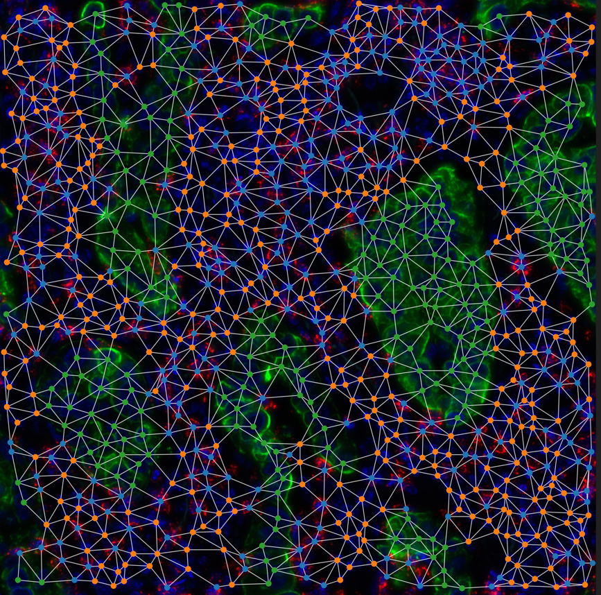
Analysis of genomic changes in each cell of a tissue during differentiation.

Key Objectives
Development of technologies based on :
- Sequencing
- Imaging
- Single-cell proteomics
Key actions
Integration of sequencing and single-cell multi-omics imaging applied to neurodevelopment and pediatric cancer:
- RNA isoform sequencing.
- Multi-omics analysis of transcriptome, epigenetic marks and chromatin and epigenome accessibility the single-cell level.
- Study of genome integrity and transcription at the level of a single cell.
Spatial imaging Development of multi-omic spatial imaging adapted to neurodevelopment :
- Signal labeling technologies, signal amplification, and clarification for multiplexed spatial imaging Staining
- Multiplexed microscopy for thick samples
- Multimodal spatial imaging
- Multidimensional spatio-temporal imaging
Proteomics for neurodevelopment and single-cell profiling of pediatric brain tumors :
- Using next-generation mass spectrometers for single-cell proteome analysis
- Development and comparison of complementary approaches
- Refining the interpretation of proteomic data with transcriptomic results
- Using experimental and analytical pipelines to characterize the origin of pediatric brain tumors
Expected results
Progress in single-cell multi-omics and sequencing techniques
- Combining single-cell multi-omics with long-read sequencing for isoform characterization
- Single-cell epigenomic multiplexing for frozen biopsies and models to map tumor epigenomic evolution
- Single-cell multi-omics to map DNA damage landscapes in relation to transcription
Improving spatial genomics and imaging technologies
- Hi-M/in situ transcriptomic imaging methods for 3D spatial information on RNA and chromatin omics
- Fast multicolor hyperspectral imaging to characterize cellular states and chromatin signatures
- New single-cell imaging modalities (RNA velocity, point mutation imaging, barcode lineage tracing imaging)
- Combining Hi-M/in situ transcriptomic imaging methods to recover 3D multi-omic spatial information
- Fast multicolor hyperspectral imaging to characterize cellular and epigenetic states
- Combining live-cell imaging with Hi-M/in situ transcriptomics to correlate individual cell lineages or phenotypes with their specific transcriptomic states
Making progress with single-cell proteomics methods
- Single-cell proteomics methods
- Bioinformatics pipeline for single-cell proteomics.
- Identifying lineage dysregulation and tumor progression using single-cell proteomics in pediatric brain tumors
This project will contribute to the development of multi-omics techniques that can be used to better understand the mechanisms of human neurodevelopment and the changes taking place in pediatric cancer models.
Les autres projets PEPR


